


Summary of findings: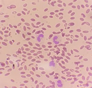
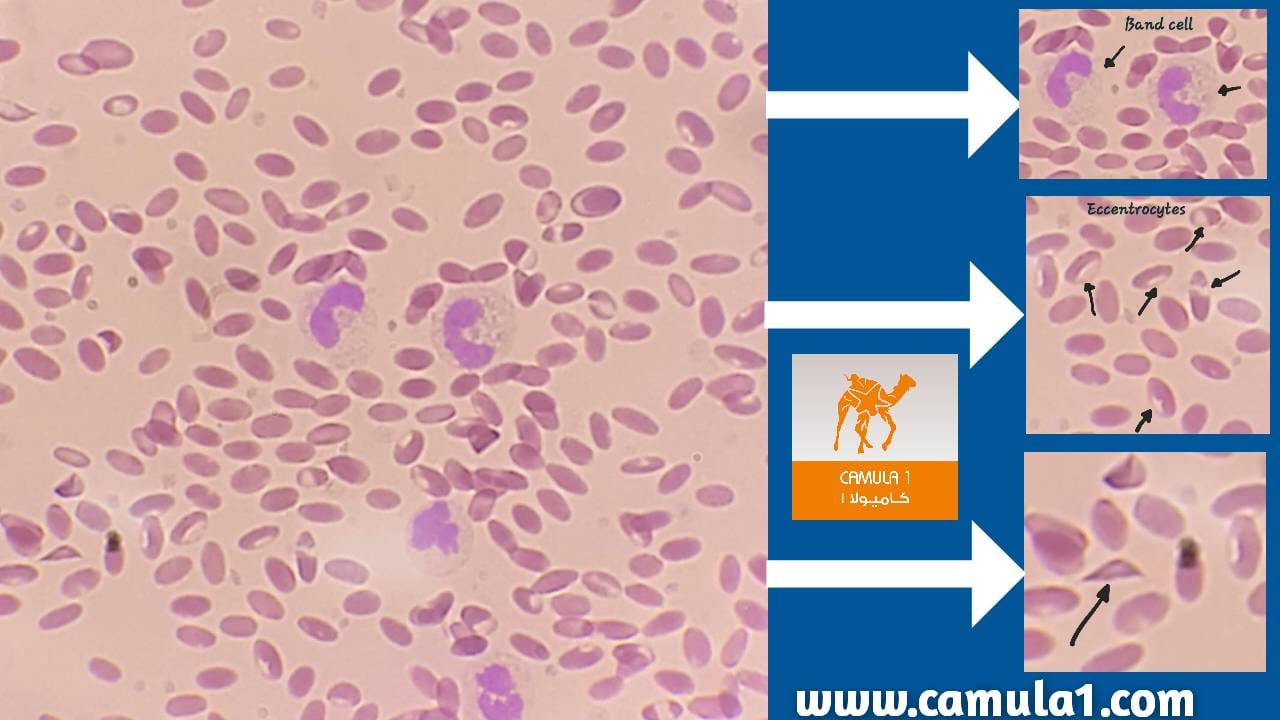
The examined peripheral blood smear demonstrates features consistent with anemia in racing camels older than five years, characterized by marked Poikilocytosis and Anisocytosis.
Morphologic details: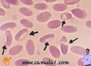
• Poikilocytosis and Anisocytosis — marked variation in red‑cell shape and size.
• Eccentrocytes — red cells with eccentric hemoglobin distribution, suggestive of oxidative membrane injury.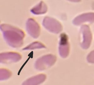
• Dacrocytes (teardrop cells) and spindle‑shaped erythrocytes (spindlocytes) — indicative of mechanical deformation or abnormal Erythropoiesis.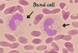
• Left shift — presence of band neutrophils consistent with an acute inflammatory response.
Differential considerations for older, high‑performance camels:
• Oxidative hemolysis (toxic or drug‑induced oxidative injury).
• Nutritional deficiencies (iron, copper, or other micronutrient deficits).
• Chronic inflammatory or infectious disease causing anemia of chronic disease or secondary marrow suppression.
• Bone‑marrow stress or dysplasia impairing effective Erythropoiesis.
• Chronic parasitism with ongoing blood loss or inflammatory burden.
• Repeated exertional or physiologic stress associated with intensive training and racing.
Clinical implications:
The combination of abnormal erythrocyte morphologies and a neutrophilic left shift supports a clinically significant anemia with concurrent inflammatory activity. These morphologic changes are not pathognomonic for a single etiology and should be interpreted in the context of clinical history, physical examination, and laboratory data.
Recommendations:
• Complete blood count to assess anemia severity and regenerative response.
• Biochemistry panel including renal and hepatic indices (BUN, creatinine, AST, LDH, GGT) to evaluate organ function and potential contributors to anemia.
• Nutritional assessment and targeted assays for iron and copper status where available.
• Parasitologic screening and infectious disease testing guided by regional prevalence and clinical suspicion.
Conclusion:
The smear indicates significant red‑cell membrane and shape abnormalities consistent with severe anemia in older racing camels. Correlation with laboratory workup and clinical context is essential to identify the underlying cause and guide appropriate management.
